Reticular tissue is a specialized form of connective tissue that dominates in areas with high cellular content. Watch the video tutorial now. White (unilocular) and brown (multilocular) fat. This renders them black and makes them easily distinguishable from type i collagen fibers that are stained red/brown. The cells that make the reticular fibers are fibroblasts called reticular cells.
Its subunits, the reticular fibers , are predominant structures in the human body, but they are mainly scattered and mixed with other types of fibers. The cells that make the reticular fibers are fibroblasts called reticular cells. Widely spaced fibroblasts are the primary cell type found in dense irregular connective tissue and they secrete proteins that assemble to form collagen. Review of loose connective tissues.
Loose connective tissue proper includes adipose tissue, areolar tissue, and reticular tissue. Widely spaced fibroblasts are the primary cell type found in dense irregular connective tissue and they secrete proteins that assemble to form collagen. Web reticular tissue is a special subtype of connective tissue that is indistinguishable during routine histological staining.
Histology Image Connective tissue

Reticular tissue, a type of loose connective tissue in which reticular fibers are the most prominent fibrous component, forms the supporting framework of the lymphoid organs (lymph nodes, spleen, tonsils), bone marrow and liver. Web.
Reticular Connective Tissue 10x Histology

Reticular connective tissue forms a scaffolding for other cells in several organs, such as lymph nodes and bone marrow. Web reticular tissue is a specific form of connective tissue predominating in several regions with high.
Reticular Connective Tissue Drawing Master the Art of Illustrating
Web in addition to the structure provided by the capsule and trabeculae, the structural support of the lymph node is provided by a series of reticular fibers laid down by fibroblasts of its reticular connective.
Web reticular tissue is a type of connective tissue proper with an extracellular matrix consisting of an interwoven network of reticular fibers that provide a strong yet somewhat flexible framework (known as the stroma) for other types of functional cells to anchor within an organ or tissue. Web reticular connective tissue, 40x. Rather, you will always find reticular cells and fibers in association with other cells. Web the major types of connective tissue are connective tissue proper, supportive tissue, and fluid tissue. Appearance and features of the reticular connective tissue.
Widely spaced fibroblasts are the primary cell type found in dense irregular connective tissue and they secrete proteins that assemble to form collagen. Web reticular tissue is a special subtype of connective tissue that is indistinguishable during routine histological staining. Comprises an abundance of reticular fibers that form complicated branching and interweaving patterns.
This Chapter Will Enable You To:
Web reticular tissue is a specific form of connective tissue predominating in several regions with high cellular content. Web the major types of connective tissue are connective tissue proper, supportive tissue, and fluid tissue. Reticular cells are specialized fibroblasts that synthesize and hold the fibers. Loose connective tissue example 2.
Web Reticular Connective Tissue 40X.
This special connective tissue forms the stroma for hemopoietic tissues and lymphoid structures and organs, except the thymus. Reticular connective tissue is named for the reticular fibers which are the main structural part of the tissue. Appearance and features of the reticular connective tissue. The cells that make the reticular fibers are fibroblasts called reticular cells.
Web Reticular Connective Tissue 10X.
Watch the video tutorial now. Web reticular tissue is a type of connective tissue proper with an extracellular matrix consisting of an interwoven network of reticular fibers that provide a strong yet somewhat flexible framework (known as the stroma) for other types of functional cells to anchor within an organ or tissue. These tissues have a peculiar feature; Connective tissue preparations are often messy with a number of blotches and shapes irrelevant to the main components of the tissue, which are the cells and the extracellular protein fibers.
These Serve To Hold Organs And Other Tissues In Place And, In The Case Of Adipose Tissue, Isolate And Store Energy Reserves.
Rather, you will always find reticular cells and fibers in association with other cells. Review of loose connective tissues. Web reticular tissue is a special subtype of connective tissue that is indistinguishable during routine histological staining. May anchor to collagenous septa, which divide organs into lobes.
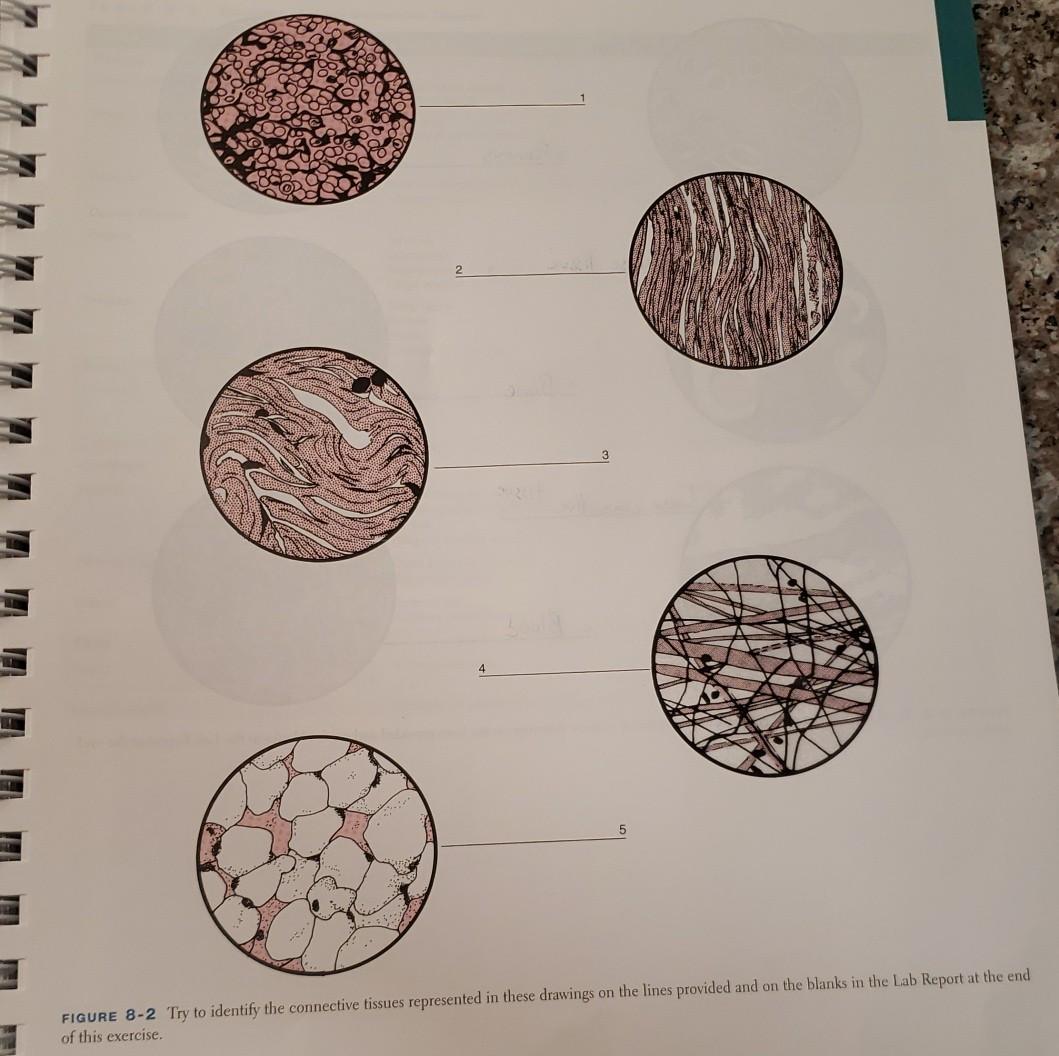
Comprises an abundance of reticular fibers that form complicated branching and interweaving patterns. Web in drawing images of connective tissue proper preparations seen under the microscope, it is important to simplify the visuals. Connective tissue preparations are often messy with a number of blotches and shapes irrelevant to the main components of the tissue, which are the cells and the extracellular protein fibers. Anatomy & physiology start typing, then use the up and down arrows to select an option from the list. Rather, you will always find reticular cells and fibers in association with other cells.